Visual Acuity Test
A visual acuity test identifies refractive errors, such as myopia, hyperopia, astigmatism, and other eye abnormalities that decrease vision clarity.
However, it does not constitute a comprehensive eye health evaluation. A normal result does not guarantee that the eyes are free of disease, as several serious conditions can develop without reduced vision. At MedPark’s Eye Care Center, a comprehensive eye exam extends beyond a visual acuity test. Leveraging advanced diagnostic technologies for in-depth assessments of ocular structures and visual quality, our ophthalmologists craft personalized plans for your optimal long-term eye health.
What is a visual acuity test?
It is an assessment of the eye’s visual performance, focusing primarily on visual clarity and sharpness, which is the eye’s ability to see and distinguish fine details, such as shapes, letters, or objects, clearly and accurately.
What does a visual acuity test assess for refractive errors?
- Myopia (Nearsightedness): Difficulty seeing distant objects clearly.
- Hyperopia (Farsightedness): Difficulty focusing on objects at close range.
- Astigmatism: An irregular curvature of the cornea that causes blurred or distorted vision.
- Presbyopia: An age-related decline in the eye’s ability to focus on nearby objects.
Common equipment for a visual acuity test
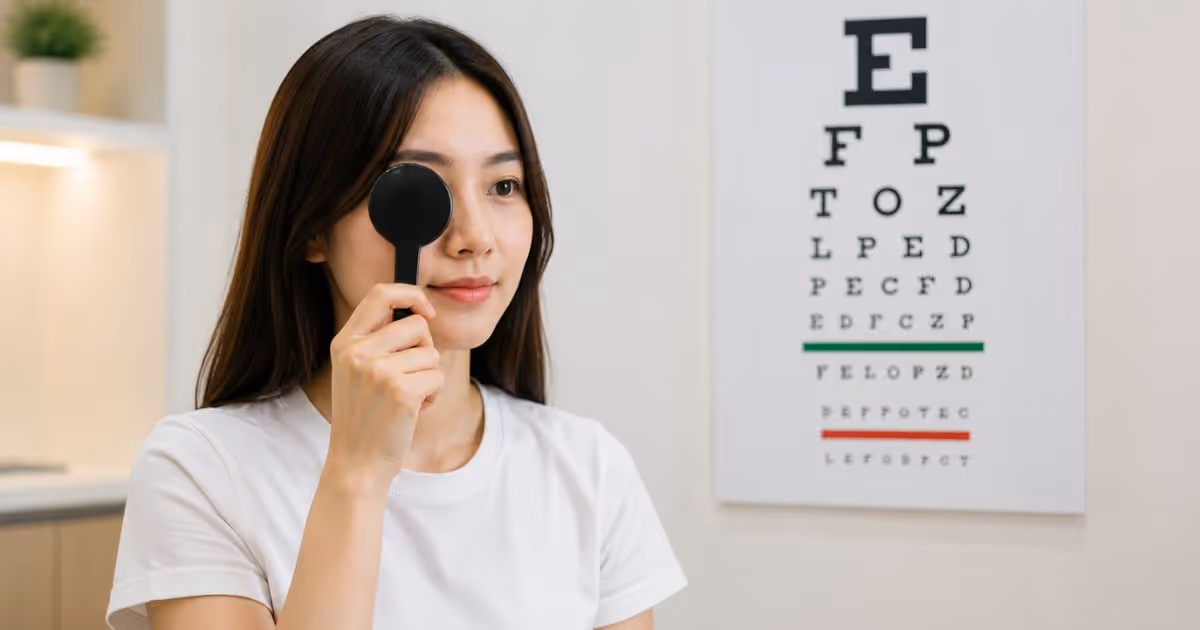
Snellen Chart
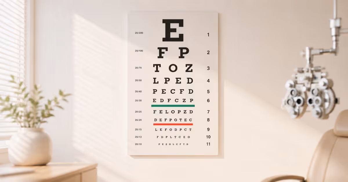
A chart or digital display that presents letters or symbols in progressively smaller sizes, used to measure distant visual acuity.
Automated refractor (Auto-Refractor)
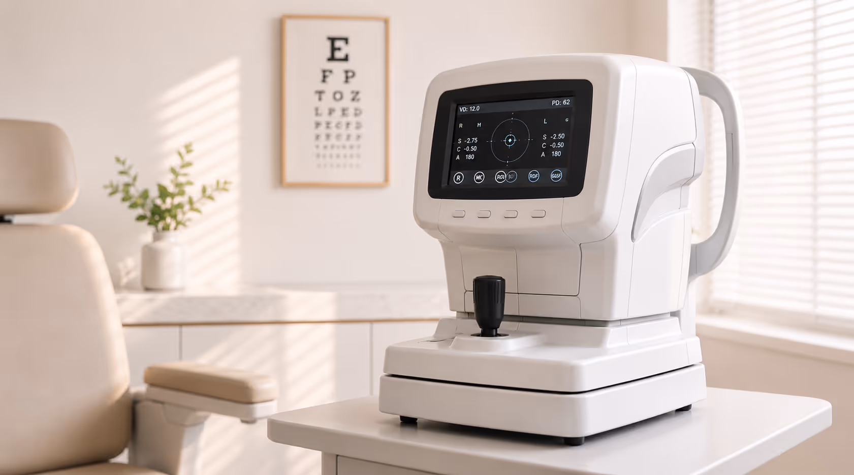
Automated refractor (Auto-Refractor) or a Retinoscope: establish a baseline value of refractive error (Objective refraction).
Slit Lamp Biomicroscopy
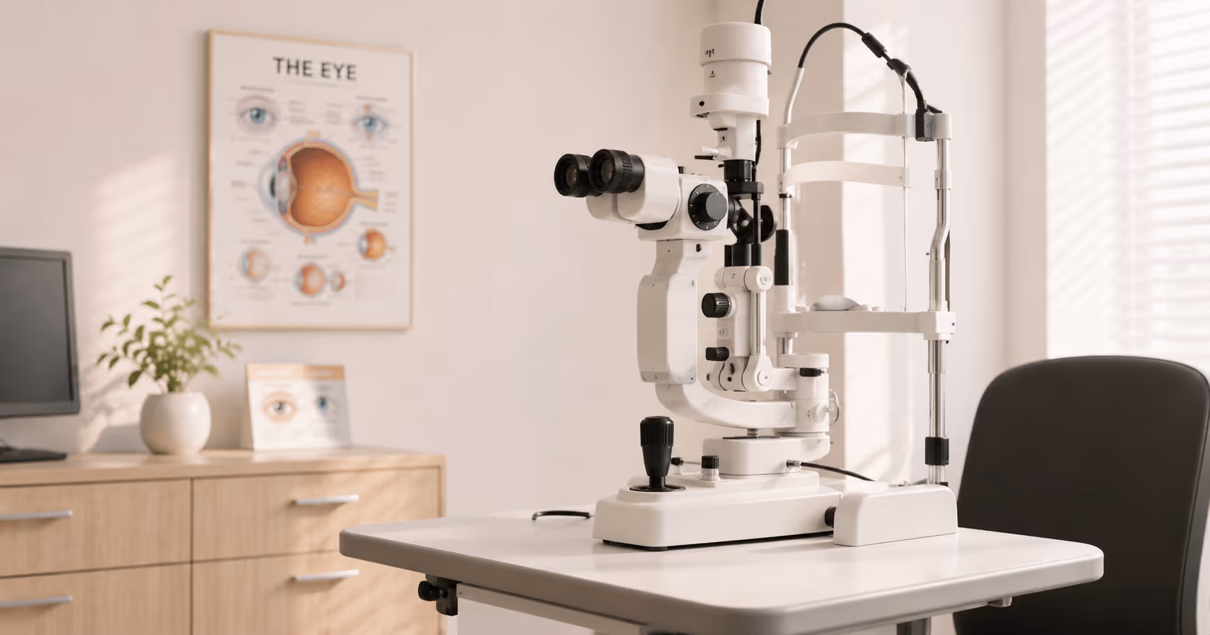
A specialized microscope for examining the anterior structures of the eye, including the cornea, lens, and iris.
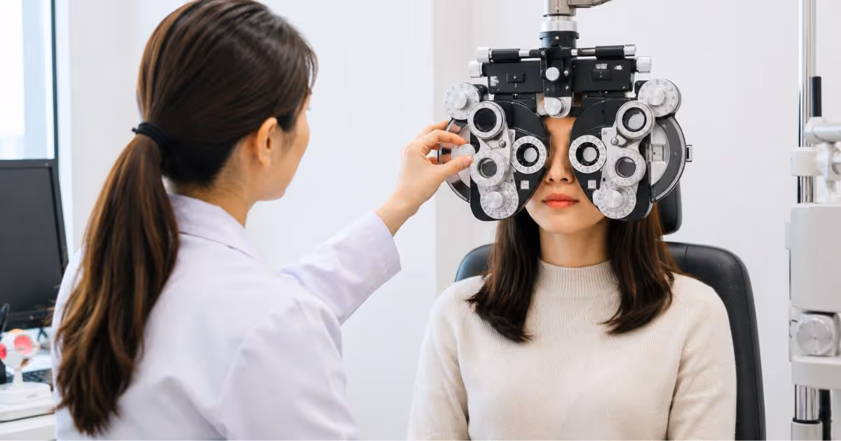
Phoropter
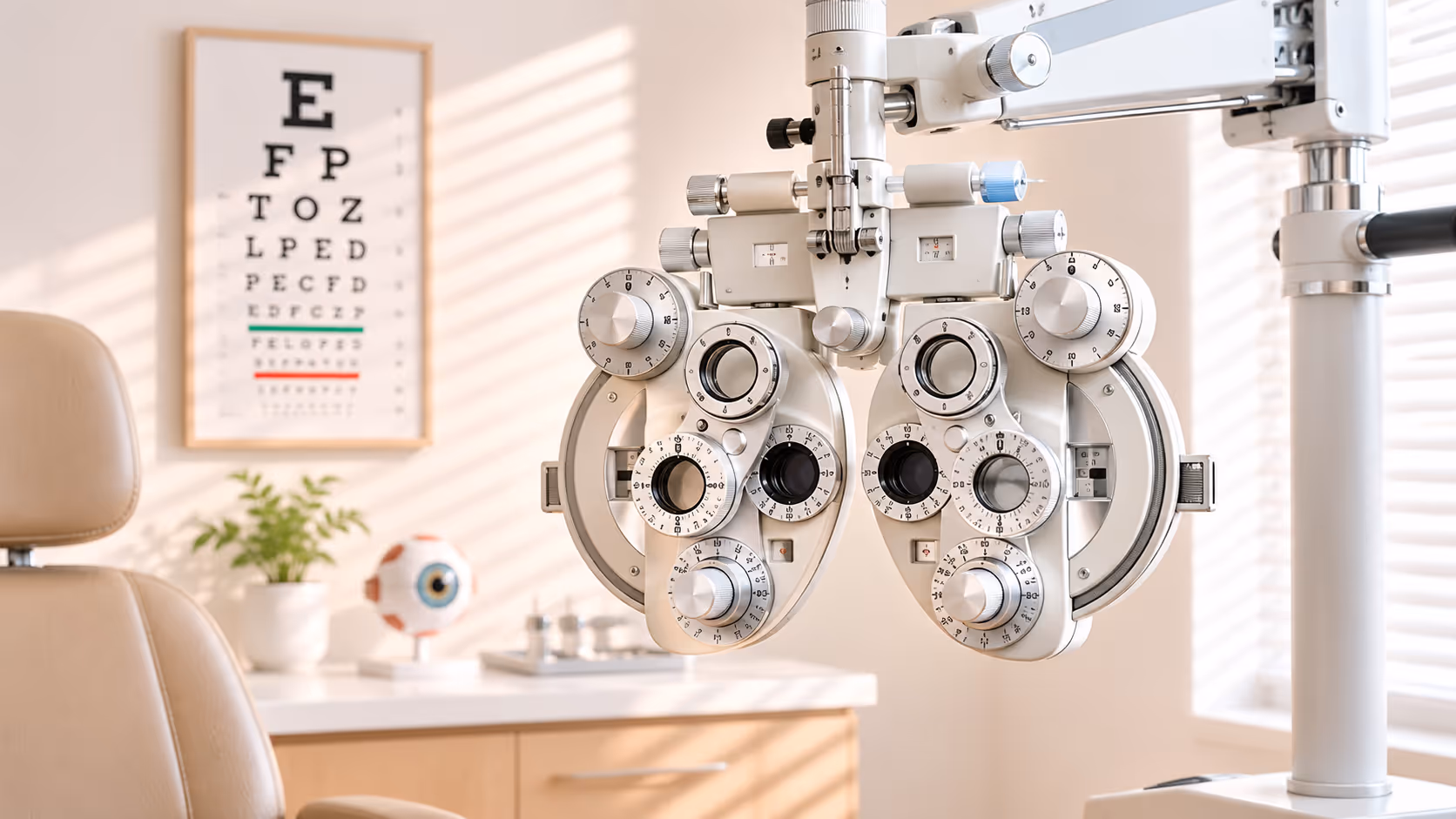
Phoropter or Trial frame and trial lens set: instruments to define subjective refraction, aimed at finding the prescription with the clearest and most comfortable vision (Subjective refraction).
Other Instruments
These may include a penlight to assess the pupillary light reflex and devices used to evaluate stereopsis (3D vision) and peripheral vision.
Results of a visual acuity test
- Visual acuity: Results are a ratio, such as 20/20. It indicates you can see details clearly at a distance of 20 feet, the standard for normal vision.
- Eye prescription: The ophthalmologist determines the precise corrective power needed for each eye. These measurements are the prescription for crafting eyeglasses or contact lenses.
- Basic eye assessment: This may include preliminary checks for conditions such as intraocular pressure and dry eye.
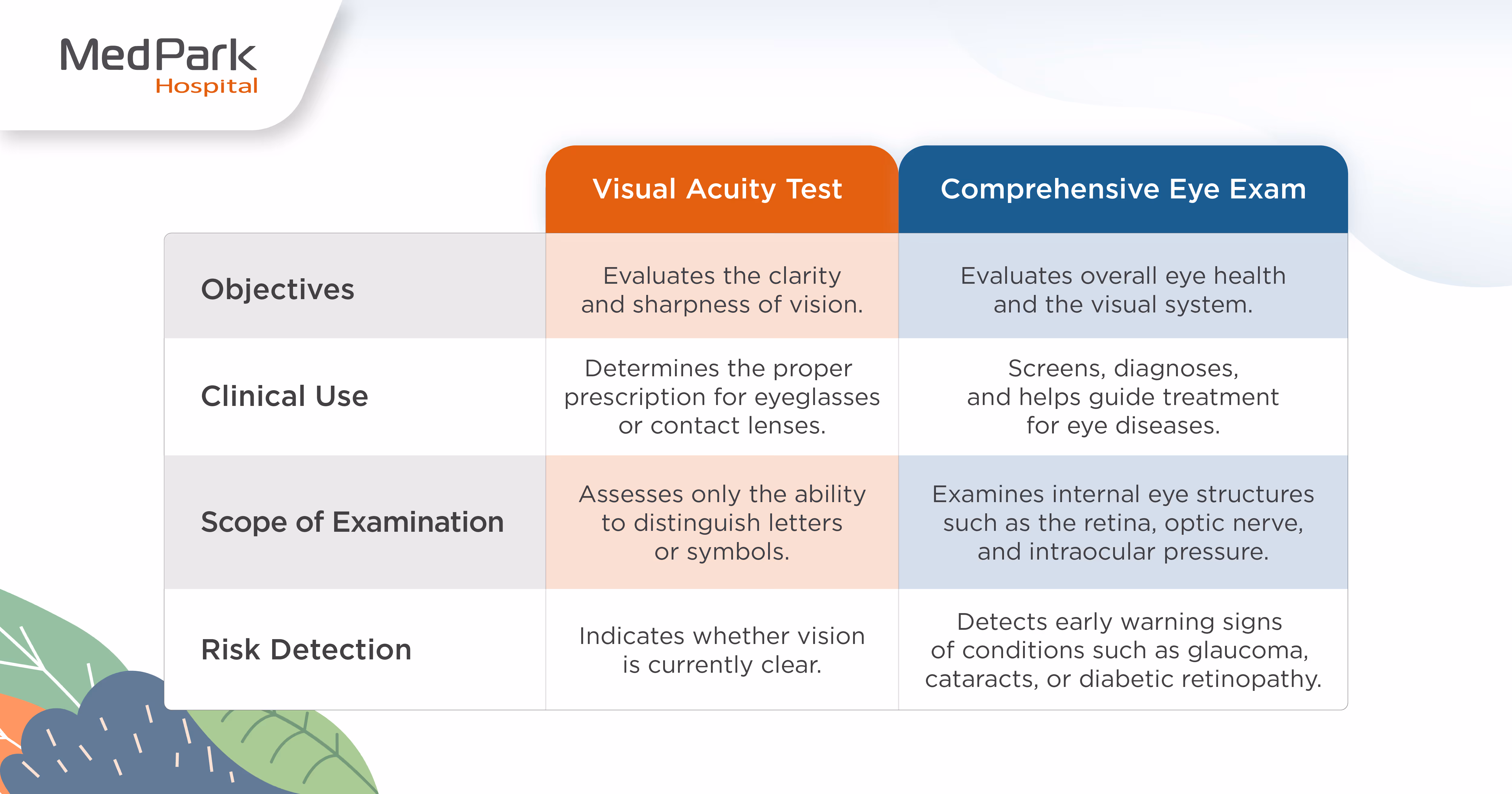
A Visual Acuity Test ≠ A Comprehensive Eye Examination. A Misconception May Affect Eye Health.
Many people assume that reading a Snellen chart clearly or having 20/20 vision means their eyes are healthy. However, a visual acuity test is only one part of a comprehensive eye examination.
A comprehensive eye examination conducted by an ophthalmologist evaluates the structures of the eye in detail to detect eye conditions that may be present but not yet manifest symptoms in their early stages.
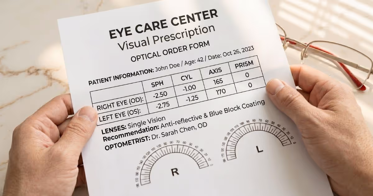
What is a visual prescription and what does it tell us?
In clinical practice, a visual prescription generally consists of two key components. The first is visual acuity, which indicates how clearly the eyes can see and distinguish fine details. The second is refraction, which measures refractive errors given as numerical values used to determine the appropriate prescription for eyeglasses or contact lenses.
Why do people with the same prescription have different visual experiences?
Even if two people have the same refractive error (for example, −2.00 diopters), the clarity and comfort of their vision may differ. Because vision involves several other factors within the eye, including:
1. Differences in eye structure
Vision does not depend solely on refractive power. It also depends on the transparency of the cornea and the crystalline lens. If early cataracts develop, visual clarity may decrease, even with glasses that have the correct prescription.
2. Retinal health
If the retina or optic nerve is affected by conditions such as glaucoma or diabetic retinopathy, vision may remain blurry even when wearing glasses or contact lenses.
3. Binocular coordination
If the two eyes are not perfectly coordinated, for instance, if they fail to focus simultaneously or if the extraocular muscles are imbalanced, this may lead to blurred vision, eye strain, or dizziness.
Limitations of a visual acuity test
- Reflects only distance-related visual clarity: The eye prescription measured by a Snellen chart only indicates central vision clarity. They cannot assess the peripheral visual field or vision, which are critical for detecting conditions like glaucoma.
- Cannot detect underlying eye diseases: Normal visual acuity does not necessarily mean the eyes are healthy. A visual acuity score alone cannot determine whether a person has elevated intraocular pressure, retinal tears, or abnormalities in the blood vessels of the eye.
- Subject to temporary fluctuations: Visual acuity can vary depending on physical conditions such as fatigue, eye strain, or certain underlying health conditions.
Visual Quality vs. Visual Acuity (Quality of Vision vs. Numerical Vision Score)
-
Visual Acuity
Refers to the ability to read letters or symbols on a Snellen chart.
-
Visual Quality
Encompasses factors that influence the overall visual experience, such as visual comfort, clarity of vision in low-light conditions, stereopsis, and the extent of the visual field.
Despite a normal eye prescription, it does not necessarily mean the eyes are free of abnormalities. A comprehensive eye exam, which includes evaluation of the eye’s key anatomical structures, helps assess both visual quality and overall eye health.
Why do vision problems persist despite a normal vision prescription?
1. Clear vision with persistent strain despite correct prescription
Headaches or eye strain at the end of the day are not always caused by an incorrect eye glass prescription. In many cases, they may be due to other factors, such as:
- Eye muscle imbalance: Misaligned eyes strain the visual system as it must exert extra effort to merge the images into a single unified view, leading to cumulative visual fatigue.
- Neurological factor: Individual variations in how the brain processes blurred images and depth of field can significantly influence visual comfort.
2. Glare and difficulty seeing at night: a possible consequence of higher-order aberrations
If your vision appears clear during the day but you notice scattered light, glare, light halos, or ghost images at night, this may be a sign of higher-order aberrations (HOAs).
- HOAs are complex refractive irregularities that are not correctable by conventional eyeglasses or standard contact lenses, and are estimated to affect approximately 15% of individuals.
- Impact on vision: HOAs often worsen when the pupils dilate in low light. As a result, visual quality may decline at night even when standard vision tests appear normal. Detecting HOAs typically requires advanced diagnostics such as wavefront technology, which identifies subtle optical imperfections in the eye.
3. Ocular surface and dry eye
Visual quality begins with the tear film, which acts as the first refractive surface that light passes through before entering the eye.
- When dry eye occurs: An unstable or irregular tear film can distort incoming light, leading to vision that appears fluctuating or temporarily blurred.
- Evaluation: An ophthalmologist may examine the eyelids, the conjunctiva, and the tear system to determine whether symptoms such as eye fatigue or blurred vision are related to dry eye disease.
4. Hidden risks in the retina and optic nerve
One of the most concerning situations is when visual acuity appears normal, yet underlying abnormalities or eye diseases may already be developing without noticeable symptoms.
- Glaucoma: This condition typically affects peripheral vision first. As a result, patients may still see clearly in the central field and be able to read letters on a Snellen chart until the disease progresses to an advanced stage.
- Optic nerve abnormalities: A detailed examination of the optic nerve head and retina, often performed after pupil dilation with an indirect ophthalmoscope or during a slit lamp biomicroscopy with a special lens, is required to determine whether the deeper structures of the eye remain healthy.
How often should you have your eyes examined?
The frequency of eye examinations depends on age, symptoms, and individual risk factors. In general, ophthalmologists recommend having a comprehensive eye examination as part of an annual health checkup to screen for eye diseases that may develop without noticeable symptoms.
Recommended eye exam frequency by age
- Children: Vision screening should begin when children start learning letters. Repeat eye examinations every 1–2 years, or sooner if a vision problem is suspected, as children’s eyes develop and change rapidly.
- Working-age adults (20–39 years): If there are no eye problems or risk factors, it is generally advisable to have at least an eye examination in the 20s and two examinations during the 30s. A more comprehensive eye examination should begin around age 40, when early age-related visual changes and the first signs of certain eye diseases may start to appear.
- Seniors (60 and older): Eye examinations are advisable every 1–2 years, or annually, depending on the advice of an ophthalmologist, as the risk of age-related eye conditions such as cataracts, glaucoma, and age-related macular degeneration increases with age.
Those who may need more frequent eye examinations
- Individuals with chronic conditions (such as diabetes or hypertension): These conditions can directly affect the blood vessels in the eyes. Patients should undergo eye examinations more frequently to monitor for complications such as diabetic retinopathy and other forms of retinal damage.
- People who use computers for prolonged periods or experience visual symptoms: If you experience end-of-day headaches, digital eye strain, blurred vision, or glare, you should consult an ophthalmologist promptly rather than waiting for your next scheduled eye examination.
- Contact lens wearers: Regular annual eye examinations are advisable to evaluate corneal health and ensure that the contact lenses remain appropriate and safe for continued usage.
Eye examination before laser or eye surgery
Before undergoing procedures such as LASIK or any eye surgeries, you should undergo a comprehensive eye examination to measure refractive errors and evaluate the structural components of the eye, including corneal curvature. After the procedures, patients should attend scheduled follow-up examinations to monitor healing and assess visual outcomes.
Why is preoperative eye examination important?
Before undergoing vision correction procedures, such as laser or eye surgery, a comprehensive eye examination is a crucial step. It helps determine whether a patient is a suitable candidate for the procedure and ensures that the treatment plan is safe and appropriate.
-
Vision assessment alone is not sufficient.
An ophthalmologist will perform a thorough evaluation of the anterior and posterior segments of the eye. These may include measuring intraocular pressure to assess the risk of glaucoma and examining the retina to detect abnormalities that could affect the safety of the procedure and its outcomes.
-
Refractive stability evaluation
The ophthalmologist will carefully review the patient’s previous prescriptions to evaluate changes in refractive error over time. Assessing refractive stability is particularly important because it confirms that the prescription is not changing rapidly or fluctuating. Stable refractive error is a key factor in ensuring the accuracy of laser vision correction or eye surgery and in achieving long-lasting results.
-
Eye structure examination
- Slit-lamp biomicroscopy: Used to assess the transparency and overall health of the cornea, iris, and lens.
- Corneal topography: A detailed scan of the corneal surface that measures its curvature and surface regularity.
- Higher-order aberration (HOA) assessment: Used to detect complex optical imperfections not correctable by conventional eyeglasses but may be addressed with advanced laser vision correction technologies.
Why can’t some people undergo eye surgery or laser vision correction?
An ophthalmologist may recommend postponing or avoiding surgery if the following conditions are detected:
- Poor ocular surface health: For example, severe dry eye, which can disrupt the tear film and affect light refraction, or the presence of corneal scarring.
- Underlying eye conditions: Such as early cataracts or abnormalities of the optic nerve. In these cases, laser vision correction may not be the most appropriate treatment.
- Structural risks of the eye: If the cornea is too thin or has abnormal curvature (for example, keratoconus), laser treatment may pose more risks than benefits.
A comprehensive evaluation by an ophthalmologist helps ensure that each patient receives the most appropriate treatment, promoting optimal visual quality and long-term eye health.
Where should you go for a reliable eye exam?
To make well-informed decisions regarding your long-term ocular health, it is essential to consult a specialized eye center equipped with advanced diagnostic technology.
Differences between an optical shop and an eye care center or hospital
- Optical shop: Primarily provides vision testing services for eyeglasses or contact lenses, typically conducted by an optometrist.
- Eye center or hospital: Offers a comprehensive eye examination conducted by an ophthalmologist, including measuring refractive error, assessing ocular alignment, examining the cornea and internal eye structures such as the retina and optic nerve, as well as screening for major eye conditions, like glaucoma, cataract, and retinal disorders. In addition, patients can receive medical advice, follow-up care, and appropriate treatment planning, including surgery or laser vision correction when necessary.

Advanced eye screening at MedPark Hospital
-
Distinguished subspecialty faculty
MedPark Hospital's Eye Care Center offers specialized ophthalmology services for cataracts, glaucoma, retinal diseases, and corneal disorders. Many of our specialists are renowned academic faculty members with over 30 years of clinical experience in Thailand and internationally. They provide expert consultations and develop personalized treatment plans tailored to each patient, particularly for complex cases.
-
Comprehensive and integrated diagnostic excellence
The Eye Care Center at MedPark Hospital is equipped with state-of-the-art technologies, including Pentacam for high-resolution Corneal Topography, Optical Coherence Tomography (OCT/OCTA) for the retina and optic nerve, and Computerized Visual Field Testing (CTVF), which are essential for identifying functional vision issues and detecting underlying eye diseases.
-
A leader in innovative vision correction
MedPark Hospital is home to the SMILE® Pro Center, featuring the latest ZEISS VISUMAX 800 technology. This advanced system enables fast, highly precise correction of myopia and astigmatism, reducing laser time to just 8–10 seconds. As a result, patients experience improved vision quality and can return to their normal activities more quickly. MedPark Hospital's Eye Care Center combines leading medical expertise with advanced technology to optimize vision and enhance quality of life.
Additional diagnostics for complex cases
In patients with complex eye conditions or those preparing for laser vision correction, information obtained from a standard vision test may not be sufficient to inform treatment decisions. In such cases, additional specialized examinations may be necessary, including:
- Corneal topography: A detailed scan of the corneal surface to evaluate its curvature and topology.
- Wavefront technology (Aberrometry): An advanced diagnostic method used to detect higher-order aberrations (HOAs)—complex optical imperfections that are not identifiable with standard vision charts and not correctable with conventional eyeglasses.
- Other ocular imaging tests: Such as optical coherence tomography (OCT) to examine the optic nerve and macula, or fundus photography to capture detailed images of the retina and detect early abnormalities.
Why is the explanation of findings important?
Going over the findings with an experienced ophthalmologist provides a clear summary of the key observations, guidance on appropriate treatment options, self-care recommendations, and a suitable follow-up plan. Moreover, you will receive an eye prescription for your eyeglasses or contact lenses if needed.
Frequently Asked Questions about the Visual Acuity Test
Is a comprehensive eye exam necessary if my vision is normal?
A visual acuity test is only one component of a complete eye exam. A comprehensive eye exam remains essential because it helps screen for eye diseases that may be asymptomatic in the early stages, such as glaucoma or retinal abnormalities, which may remain undetected without a detailed examination.
Is the visual acuity test painful?
It is not painful. In some steps, such as measuring eye pressure with a non-contact tonometer, you may feel a brief startle from a puff of air directed at the surface of the eye. The test is quick, less than a split second.
Can my vision prescriptions change over time?
Yes. Vision prescriptions change over time due to age and other health-related factors. In children, the eyes are still developing, so vision may change more frequently. Regular eye examinations are therefore advisable. In adults, noticeable vision changes often begin around the age of 40, including the development of presbyopia (age-related farsightedness). The risk of certain eye diseases may also increase during this period. For this reason, a baseline eye exam at age 40 is strongly advisable.
Can I have my vision tested without getting glasses immediately?
Yes. After the examination, the ophthalmologist will provide a prescription, which you may use to obtain glasses at your convenience. However, it is generally advisable to obtain glasses within 3–6 months, as vision may change over time, especially in children, adolescents, or individuals whose vision has not yet stabilized. If more time has passed or if you experience blurred vision, eye strain, or noticeable changes in your vision, you should undergo another eye examination to ensure an accurate prescription.
Can a visual acuity test detect eye diseases?
The Snellen chart measures only the sharpness of vision and can identify refractive errors. To determine whether an eye condition is present, a comprehensive eye examination is necessary.
How long does an eye exam take?
A comprehensive eye examination, including vision testing, typically takes 45 to 90 minutes. The duration may vary depending on an individual's eye condition and whether additional procedures are required, such as pupil dilation to allow a more detailed examination of the retina.
What are the side effects of pupil dilation?
After pupil dilation, some people may experience temporary blurred vision and increased sensitivity to light, as the pupils remain dilated. These effects usually resolve on their own within 4-6 hours. It is advisable to bring sunglasses and, if possible, have someone accompany you and drive you home after the examination.
Who should have their eyes examined more frequently?
Individuals at higher risk, including those 60 or older, people with diabetes or hypertension, individuals with a family history of eye disease in first-degree relatives, contact lens wearers, and those with a history of eye surgery or ocular trauma, should undergo eye examination as advised by an ophthalmologist.
Summary
A visual acuity test is a fundamental examination used to evaluate the sharpness of vision and the ability to distinguish fine details of objects at various distances. The test typically involves using a Snellen chart, together with refractive assessment tools such as a phoropter or trial frame and trial lens set, to measure refractive error. This examination helps diagnose common vision conditions, including myopia (nearsightedness), hyperopia (farsightedness), astigmatism, and presbyopia. The results are usually expressed in numerical form, such as 20/20, and are used to determine the most appropriate prescription for eyeglasses or contact lenses tailored to each individual.